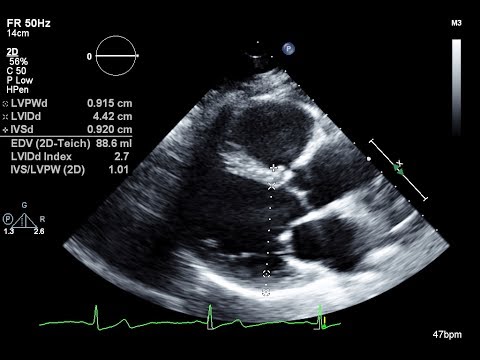
2D Echocardiography or 2D Echo is a heart test in which an ultrasound technique is used to take pictures of the heart. It shows a cross-sectional view of the beating heart, chambers, valves and the major blood vessels of the heart. Doppler is a special element of the 2D Echo test that assesses blood flow in the heart. Cardiologist recommends a 2D Echo test for people who experience abnormal pain in the chest area, breathlessness, blood pressure etc.
How is 2D Echo done?
A 2D Echo test is done by a cardiologist. A colourless gel is applied to the chest area. The patient is asked to sleep. The cardiologist moves the transducer across the various parts of the chest to get specific/desired views of the heart.
The cardiologist gives instructions to the patient such as breathing slowly or holding breath. This helps in getting superior-quality images of heart functions. The images are viewed on the monitor and recorded on paper, video or DVD. The cardiologist later assesses and analyzes the recordings deeply.


